What conditions can it benefit?
An ultrasound scanner provides the opportunity to assess almost any tissue in real time, except for organs or tissues that are very deep or difficult to access; the rest can be assessed. Conditions that can benefit from the services of an ultrasound scanner:
- Tendon injuries: tendonitis or tendinopathies. The ultrasound can show how the tendon fibres are located and whether there is any damage or whether everything is in perfect condition.
- Muscle and ligament injuries: strains or tears. It is possible to visualise the condition of the muscle/ligament fibres and whether it is related to the discomfort you are suffering.
- Nerve injuries: nerve entrapment or thickening. Conditions such as carpal tunnel, sciatic nerve entrapment or other nerves in the body can be assessed by ultrasound and their route from the spinal column to other territories can be analysed; it is possible to see where the problem area is located and treat it.
- Inflammation in joints due to inflammatory processes such as bursitis, or due to traumatic events such as a fall or accident. In this case, the ultrasound scanner can be used to observe the accumulation of fluid in the injured area, allowing us to be very specific and helping to work on the specific area.
- Small superficial bone fractures or joint wear and tear (arthrosis). With an ultrasound scan you can find out if a specific area suffers from excessive wear and tear (arthrosis) or if there is even a bone fracture.
- Muscle condition. Sedentary people, who do not do much physical activity, with high levels of stress or who do not even follow a balanced diet, may have a greater amount of infiltrated fat in their muscles, indicating that the musculature is weak and in poor functional condition.
How can progress with treatment be seen?
Thanks to the latest technology that Madrid Health offers, it is possible to create a specific treatment plan based on the results of the ultrasound test. Progress will depend on the degree of tissue involvement and it usually takes several weeks to see significant changes:
- If there is elongation, rupture, thickening or degeneration of a tendon, ligament, nerve, bone or muscle, it is possible to see the healing and regeneration process a few weeks after the first ultrasound, comparing the results with the next ultrasound and measuring the quality of the tissues, thus proposing the best plan of action at any given time.
- In inflammatory processes, it can be observed how the inflammation is considerably reduced in the affected area, improving the condition and reducing the pressure in the area and the discomfort it was causing.
- One of the most important aspects of Madrid Health's treatment plans is an exercise rehabilitation process that is carried out through Physitrack. Exercise is fundamental to any injury, so the professionals at Madrid Health will selectively choose exercises based on each patient's condition. If the patient does them frequently, follows the healthy habits indicated and keeps active, it is possible to see the evolution of their muscles and the amount of infiltrated fat with the ultrasound, which will be less the more the patient moves and the higher the level of physical activity they practice.
What do you receive at an ultrasound session?
Apart from the latest technology, the knowledge that the professionals at Madrid Health have acquired over many years of practice and continuous training, exquisite treatment and care for each and every one of their patients. In addition to all this, the ultrasound session includes:
1. A complete ultrasound examination of the affected area in real time with feedback from the professional.
2. Oral explanation of the procedure at all times.
3. Photo and video signage of the scanned area.
4. Written ultrasound report specifically detailing and describing the state of the structures that have been analysed.
5. Photos taken during the examination so that the patient can see the area studied for themselves.
Template:
- What is an ultrasound scanner? It is a health, anatomical and functional tool that helps to assess the state of certain structures of the human body: muscles, tendons, nerves, ligaments, bones, joints and organs. The ultrasound scanner makes it possible to see and assess in real time how the tissue being studied is doing.
- What have we found?
- Recommendations: another ultrasound study in X month/months
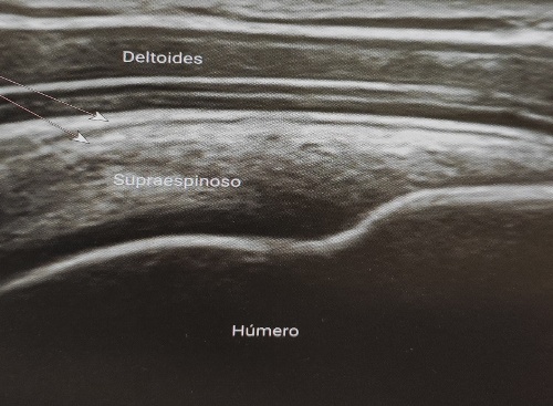
Below is a healthy ultrasound image of the supraspinatus tendon of the shoulder, the most affected tendon in shoulder injuries and part of the rotator cuff. As can be seen, the supraspinatus tendon is located between 2 structures: superior to it is covered by the deltoid muscle and inferior to it is the humerus bone, where the tendon inserts.
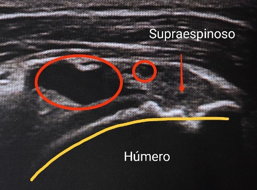
However, the following ultrasound image shows pathology. It is another supraspinatus tendon where the same structures appear as in the previous image. Let us analyse in detail by means of a physiotherapy diagnosis, which analyses the state of the structures, what the differences are between a healthy image and an altered one:
1. The cortical line of the humerus bone has been altered, it is not linear as it should be in a healthy bone.
2. The thickness of the supraspinatus tendon is greater than a normal tendon.
3. The shape of the supraspinatus tendon is altered and does not correspond to a normal one.
4. The fibrillar pattern of the supraspinatus tendon is altered, with dark areas that may indicate inflammation and/or separation of its tendon fibres.
Madrid Health is pleased to present its latest acquisition in technological innovation: a portable ultrasound scanner. An ultrasound scanner is a health, anatomical and functional tool that helps to evaluate the state of certain structures of the human body: muscles, tendons, nerves, ligaments, bones, joints and organs.
The ultrasound scanner makes it possible to see and assess in real time how the tissue being studied is in order to be able to make a physiotherapy diagnosis. This type of diagnosis is slightly different from medical diagnosis, let's look at the differences:
1. The medical diagnosis gives a name to the state of the tissue being studied, for example: tendinitis, ligament rupture, bursitis, fracture, etc.
2. Physiotherapy diagnosis does not give names, it is not its function, it only describes the state of the tissues, for example: instead of tendonitis, the diagnosis would be inflammation of the tendon; instead of ligament rupture, the diagnosis would be loss of continuity in the union of the ligament fibres.
3. Which is better? Neither one nor the other, both are correct and want to help to know the state of the patient's structures.
Apart from being able to provide a very specific diagnosis and to know with great certainty what is happening in the tissue, the use of ultrasound has multiple other benefits that Madrid Health would like to share with you:
- Both the diagnosis and the ultrasound scan are carried out in a short space of time and it is not necessary to wait weeks or months to be able to receive this type of service.
- It is a safe, non-invasive, painless approach that does not produce side effects on patients' health.
- Not only does it help to provide a diagnosis to the patient, but it can also assess the evolution of the tissues some time after the first diagnosis; thus seeing how the lesion has progressed and whether the progress has been positive or not.
- Ultrasound is an ultrasound that works with sound waves, which do not produce ionising radiation like X-rays.
- This diagnostic technique makes it possible to take photographs and videos instantly.
- It is possible to use the ultrasound scanner as an aid when performing other techniques such as shock waves, dry needling or neuromodulation. It provides information and safety in the technique.


