
How do you know which tendon is affected? Ultrasound may be the answer
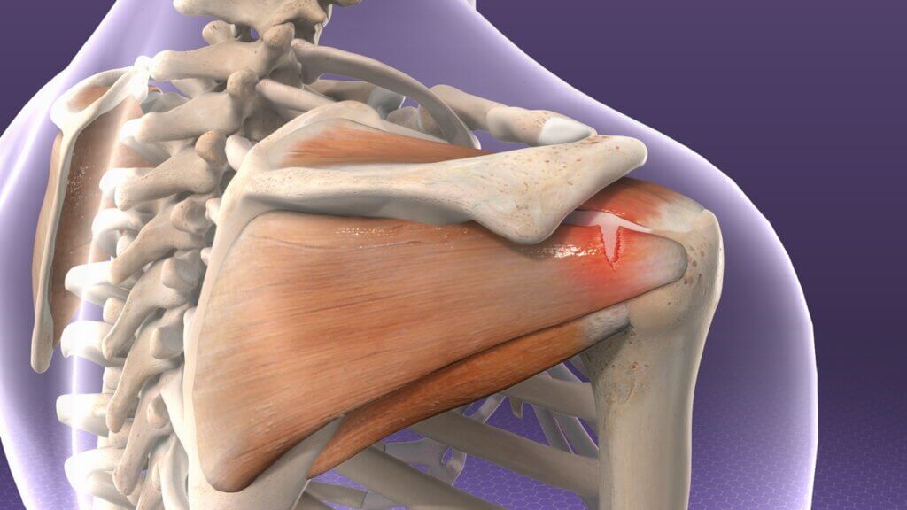
The shoulder, or better known as the shoulder girdle, is a set of anatomical structures with five joints and their corresponding tissues. These joints are: glenohumeral, subdeltoid, scapulothoracic, external-clavicular and acromion-clavicular. Each is protected by numerous ligaments, tendons, bursae and muscles, as well as other tissues such as skin, subcutaneous tissue, nerves and bones. This makes the shoulder the most mobile joint in the human body.
However, there is a biomechanical handicap: the greater the mobility, the greater the wear and tear on these structures. And here comes the main question: when a person suffers from shoulder pain, which structure can be the cause of the discomfort or be affected? Therapists and doctors have extensive knowledge of anatomy, biomechanics and orthopaedic and neuromuscular tests that help to know which movements are painful and which tissues may be affected.
However, in the shoulder girdle it is difficult and complex to know which structure causes the problem, as the space where all the tissues congregate is small and the number of tissues is vast. Everything is interrelated and if any tendon, ligament or muscle is inflamed and/or degenerated it will produce irritation in the nearby structures, so it is very difficult to find out by palpation or orthopaedic and neuromuscular tests which structure is affected.
So, what can we do to find out and get an answer to this problem? An imaging test that assesses the state of all tissues in real time both in movement and in static. That test is ultrasound: it is painless and non-invasive, has no side effects, shows results in real time and allows photos and videos to be taken. Ultrasound shows high sensitivity and specificity in its results, making it an ideal imaging test when assessing the shoulder.
The ultrasound assessment protocol divides the shoulder into different body zones and analyses all structures one by one, allowing the exact status of all structures to be known and providing essential information. The injured shoulder and the healthy shoulder should always be analysed to see if there are significant differences between the two. Once the ultrasound results are obtained, the person in charge of performing the test needs to thoroughly evaluate all the photos and videos that have been saved; looking for alterations in the tissues and comparing both shoulders.
After completing this visual examination, an ultrasound report is drawn up detailing the type of evaluation performed and the anatomical findings with the alterations found and where. By knowing what type of tissue is altered, it is possible to make a more specific treatment plan focused on the recovery of that structure. In this way, a better state of well-being can be achieved and the recovery process will be better and faster.