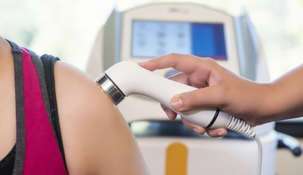
An ultrasound scanner is a health, anatomical and functional tool that helps to assess the state of certain structures of the human body: muscles, tendons, nerves, ligaments, bones, joints and organs. The ultrasound scanner makes it possible to see and assess in real time the state of the tissue being studied and thus be able to make a physiotherapy diagnosis.
Apart from being able to provide a very specific diagnosis and to know with great certainty what is happening in that tissue, the use of ultrasound has multiple other benefits:
- The whole process is carried out instantaneously and in real time, with no need to wait. It is possible to take photos and videos.
- It is a safe, non-invasive, painless approach that does not produce side effects on patients’ health like X-rays.
- Not only does it help to provide a diagnosis to the patient, but it can also assess the evolution of the tissues some time after the first diagnosis; thus seeing how the lesion has progressed and whether the progress has been positive or not.
- It is possible to use the ultrasound scanner as an aid when performing other techniques such as shock waves, dry needling or neuromodulation. It provides information and safety in the technique.
As can be seen, the ultrasound scanner is a very versatile tool that provides both immediate diagnostic information as well as help in locating the area to be treated. There are two types of ultrasound scanners today:
1) Fixed. Tethered to a computer and printer with cables. They are heavy, take up a lot of space but offer superior image quality.
2) Portable. With or without cables, working by Wifi. They are light, easy to carry and very practical, however the battery life is short and the image quality is lower compared to the fixed ones.


